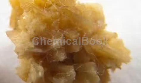
Urine is a complex physical and chemical system. The formation of urinary tract stones is the process of converting liquid substances in urine into solid substances. This process requires a certain amount of energy, and urinary supersaturation caused by high concentrations of stone-forming substances in the urine is the energy source that drives stone formation. The formation of stones depends on the chemical potential difference between the liquid and solid phases. When urine is saturated, the liquid phase tends to transform into the solid phase. The formation of uroliths is not caused by a single factor, but is the result of a combination of factors. In the process of stone formation, although urinary supersaturation is an important prerequisite, it is sometimes not necessarily the only condition. Supersaturation often requires the participation of other factors to form stones, especially the balance between urinary saturation and crystallization inhibitory factors. Under normal circumstances, the saturation of certain stone-forming substances in urine often exceeds its solubility.
Clinically, most stones are calcium oxalate stones. Calcium oxalate stones may be a polygenic hereditary disease. Genes can influence stone formation by regulating calcium, oxalate, and citrate. The concentration of calcium oxalate in normal urine is 4 times its solubility, and precipitation will only occur when the concentration of calcium oxalate reaches 10 times its solubility. This mainly depends on the activity of the crystallization inhibitory factor, which can adsorb At the growth point on the crystal surface, it prevents the nucleation, growth and aggregation of crystals. Crystallization inhibitory factors can also combine with certain stone-forming substances to form soluble conjugates and reduce the urinary saturation of these stone-forming substances. Clinically, although some people have increased urinary excretion of oxalic acid and calcium, they may not necessarily form stones. This is also due to the effect of crystal inhibitory factors in the urine. Some common important inhibitors are citrate, pyrophosphate, and magnesium. At the same time, the reduced content of these inhibitory factors in urine is also an important condition for stone formation.

Other substances that contribute to the formation of uroliths include crystallization-promoting factors in urine, but they are not as important as crystallization-inhibiting factors. Simple promoters are rare. Certain substances in urine can play a dual role in promoting and inhibiting crystal formation at different stages. For example, glycosaminoglycan promotes crystal nucleation but inhibits crystal aggregation and growth. Stones are mainly composed of crystals, so the stone formation process also follows the chemical kinetics of crystal formation. This process roughly goes through the following steps: crystal nucleation → crystal growth → crystal aggregation → crystal retention → stone formation.
The direct causes of calcium oxalate stone formation include hypercalciuria, hyperoxaluria, hyperuricosuria and hypocitrateuria.
1. Hypercalciuria:
Hypercalciuria is defined as urinary calcium excretion >200 mg/day or >4 mg/kg/day under random diet. Among calcium oxalate stones, hypercalciuria is the most common metabolic disorder, accounting for approximately 30%-60%. Calcium is mainly absorbed in the small intestine, filtered by the kidneys, and reabsorbed from the renal tubules. Parathyroid hormone (PTH) and 1,25-dihydroxyvitamin D are involved in regulating the calcium balance in the body. The organs they regulate include the kidneys, intestines If the regulatory functions of these organs are abnormal, it will lead to calcium metabolism disorders. There are three main types of hypercalciuria: ① Absorptive hypercalciuria, caused by excessive intestinal absorption of calcium; ② Renal hypercalciuria, caused by reduced reabsorption of urinary calcium by the kidneys; ③ Reabsorptive hypercalciuria Hypercalciuria is caused by increased mobilization of calcium from bones.
2. Hyperoxaluria:
Hyperoxaluria refers to the excretion of oxalate in urine >45mg/day. About 80% of the oxalic acid in the human body is the end product of intrahepatic synthesis and vitamin C metabolism. The rest comes from oxalic acid in food, which is absorbed in the stomach, small intestine and colon and excreted by the kidneys. In urine, oxalic acid increases calcium oxalate saturation 10 times more than calcium. If the urinary oxalic acid concentration increases by 10% from the daily excretion of 45 mg to 49.5 mg, it is equivalent to a 100% increase in urinary calcium, which is equivalent to an increase in the daily urinary calcium excretion from 200 mg to 400 mg. Therefore, increased urinary oxalic acid excretion is a more dangerous stone-forming factor. There are three main types of hyperoxaluria: ① primary hyperoxaluria, caused by excessive endogenous oxalic acid production; ② enterogenic hyperoxaluria, caused by excessive absorption of exogenous oxalic acid; ③ idiopathic hyperoxaluria Hyperoxaluria, the cause is unknown, may be related to the enhanced function of red blood cells in transporting oxalate.
3. Hyperuricemia:
Hyperuricosuria refers to the excretion of uric acid in urine >600mg/day. Clinically, about 15% of calcium oxalate stones are caused by hyperuricosuria. The main cause of hyperuricemia is excessive protein intake; secondly, excessive uric acid synthesis in the body, which cannot be corrected even if protein intake is restricted. Calcium oxalate stones caused by hyperuricosuria are called hyperuricosuric nephrolithiasis (HUCN). The formation process of HUCN has been basically elucidated. It is the formation of calcium oxalate stones induced by sodium urate through the orientation epiphysis mechanism. When the urine pH value is >5.5, supersaturated uric acid dissociates in sodium-containing urine and forms sodium urate. After sodium urate precipitates and crystallizes, it directly induces the formation of calcium oxalate crystals through heterogeneous nucleation. Excessive sodium urate in urine can also combine with certain calcium oxalate crystal inhibitory factors in urine, thereby indirectly promoting the formation of calcium oxalate crystals.
4. Hypocitrateuria:
In calcium-containing stones, low citrateIt is mediated by parathyroid gland cell membrane-bound adenylyl cyclase. This enzyme requires Mg2+ to activate, and at this time, the plasma Mg2+ concentration decreases, so it is difficult to activate this enzyme. Therefore, although blood calcium has initially decreased, it cannot stimulate the parathyroid glands to secrete PTH, and blood calcium further decreases, leading to hypocalcemia. At this time, the response of PTH target organs such as the skeletal system and renal tubular epithelium to PTH also weakens. This is because PTH must also be mediated through adenylyl cyclase to promote the functional activities of target organs. In hypomagnesemia, adenylyl cyclase on target organs cannot be activated either, so bone calcium mobilization and calcium reabsorption in the renal tubules are hindered, and blood calcium cannot be replenished.
Many experimental studies have shown that administration of magnesium salts can prevent stone disease. Mice fed a pyridoxine-free diet are susceptible to stone formation, which can be prevented by supplementing magnesium. Another example is the administration of hexanediol to rats, which can cause calcium oxalate deposition in the kidneys only in the second half of a normal diet. However, in a magnesium-deficient diet, calcium deposition occurs within 24 hours. Some scholars have found that areas where the soil is deficient in magnesium have a higher incidence of stones, and the urinary magnesium/calcium ratio of stone patients is lower. The “stone belt” of the southeastern United States generally coincides with areas of magnesium-deficient soil. A large sample survey from Dallas, USA found that the detection rate of low magnesium calcium stones was 4.3%.
By far the most common cause of marked hypomagnesia is inflammatory bowel disease associated with malabsorption. Most patients with hypomagnesuria also have hypocitraturia. In these individuals, loss of the inhibitory or binding ability of magnesium or/and citrate is associated with calcium oxalate crystal production. Several clinical trials have examined the effectiveness of magnesium in preventing recurrence of stones. In addition, a comparative study of magnesium metabolism in urinary stone patients and normal controls found no magnesium deficiency or negative magnesium balance in the diet of stone patients. Although some studies have found low urinary magnesium excretion in kidney stones, stones can form even with normal urinary magnesium levels. The reduced urinary magnesium/calcium ratio in patients with stones is related to increased urinary calcium excretion, rather than being the result of reduced urinary magnesium excretion. A special disease. Because the protein encoded by Claudin-16 (CLDN16) is a tight-binding protein, its mutation can cause familial hypomagnesemia accompanied by hypercalciuria and nephrocalcinosis, which can ultimately lead to renal failure. exhaustion. The main reason for this change is that TAL deficiency in the loop of Henle leads to disorders of renal tubular calcium and magnesium reabsorption. The CLDN16 heterozygous form may have milder symptoms and delay the process of kidney damage. However, the specific role of this gene polymorphism in idiopathic hypercalciuria has not yet been studied in depth.

 微信扫一扫打赏

